An observation that cannot be categorized as LI-RADS 1 2 4 5 because of equivocal imaging features. Flow imaging of renal arteries was carried out to quantify the dependence of renal blood flow on the cardiac cycle.

Impact Of The Sampling Rate Of Dynamic Myocardial Computed Tomography Perfusion On The Quantitative Assessment Of Myocardial Blood Flow Clinical Imaging
Ad Laser Doppler Blood Flow Assessment Clinical and Research Application.

. Insonation of targeted microbubbles produces regions of reduced blood flow within tumor vasculature. Lee Jason Ng Maria Carr James Carr Jeffrey J. A camera will focus on the heart for approximately 10 minutes and measure the amount of imaging material that.
Blood flow velocity measurement with phase contrast magnetic resonance imaging PC-MRI is widely applied in clinical routine imaging. Purpose To investigate the feasibility of blood flow imaging in the parotid gland using the arterial spin labeling ASL technique for assessment of functional changes in. Magnetic resonance imaging MRI allows the noninvasive assessment of absolute myocardial blood flow MBF in mLming during rest and hyperemia induced by intravenous adenosine a powerful coronary vasodilator with short decay time.
4D flow magnetic resonance imaging measured in vivo 3D blood flow velocities in 60 AF patients and 15 controls. An observation that meets criteria for LI-RADS 4 or 5 with stable imaging features for 2 years AND no increase in diameter for 2 years. What is the difference between laminar flow turbulent flow and vortex flow.
Because absolute MBF thresholds may be affected by technique methodology and the microvasculature we investigated whether a relative measure of MBF improves accuracy to identify hemodynamically significant coronary stenosis. Absolute values of blood flow and arterial transit times were determined in the kidney cortex. A total of 143 participants in the Multiethnic Study of Atherosclerosis COPD Study were examined by dynamic contrast-enhanced pulmonary perfusion magnetic resonance imaging MRI at 15 T.
How is blood flow measured in a vessel. Preliminary experimental results are encouraging and this imaging modality may prove clinically feasible for detecting and monitoring the early antitumor effects in response to cancer drug therapy. Quantitative myocardial perfusion imaging by computed tomography CT was recently introduced to calculate myocardial blood flow MBF.
During the examination you will be asked to rest on a scanning table. What are time-of-flight effects. What are the usual velocities of blood found in the human vascular system.
Left atrial 4-dimensional flow magnetic resonance imaging stasis and velocity mapping in patients with atrial fibrillation Michael Markl Daniel C. ECG-triggered DWI was acquired in the coronal plane with 16 b values in the range of 0 smm2 and 750 smm2 at the time of minimum MIN 20 milliseconds after R wave and maximum renal blood flow MAX 197 24 milliseconds. The quantitative values were in good agreement with the reference values obtained after calibration of the total flow.
Ad Heart Imaging Technologies is now part of Intelerad Medical Systems. High-pressure fragmentation of microbubbles bound to tumor endothelial receptors reduced blood flow within 2 syngeneic mouse tumor models for 30 minutes. VCEUS imaging was shown to be a promising modality for monitoring changes in tumor blood flow.
Anatomic maps of LA and LAA stasis and velocity were calculated to quantify atrial peak velocity mean velocity and stasis velocities. Usually velocity and volumetric flow measurements are performed using unidirectional encoding of the through-plane velocity with a 2-dimensional 2D acquisition. Isolated organ hemoperfusion allows validating perfusion imaging.
Quantitative myocardial perfusion imaging by computed tomography CT was recently introduced to calculate myocardial blood flow MBF. Pulmonary microvascular blood flow PBF was calculated on a pixel-by-pixel basis by using a dual-bolus technique and the Fermi. Preinjection of an anti-CD41 antibody blocked the reduction of tumor blood flow where a reduction in blood flow was observed in only 1 of 26 animals.
Because absolute MBF thresholds may be affected by technique methodology and the microvasculature we investigated whether a relative measure of MBF improves accuracy to identify hemodynamically significant coronary stenosis. Leader in medical imaging management software specializing in diagnostic viewing. Discover all of the pertinent medical information regarding magnetic resonance imaging.
Hu X1 Kheirolomoom A Mahakian LM Beegle JR Kruse DE Lam KS Ferrara KW. Ad Learn the uses and side effects of magnetic resonance imaging MRI scans today. How can you predict whether a certain vessel will be bright or dark on an MR image.
What are spin-phase effects. The greatest difference observed was 13. Imaging uses very small doses of a radioactive imaging agent that is injected into the blood stream.

Molecular And Nonmolecular Magnetic Resonance Coronary And Carotid Imaging Arteriosclerosis Thrombosis And Vascular Biology

Can 3d Pseudo Continuous Arterial Spin Labeling Perfusion Imaging Be Applied To Predict Early Response To Chemoradiotherapy In Patients With Advanced Nasopharyngeal Carcinoma Radiotherapy And Oncology
Imaging Methods For Surgical Revascularization In Patients With Moyamoya Disease An Updated Review Springerlink

Pin On Nurse Stuff
Frontiers Coronary Magnetic Resonance Angiography In Chronic Coronary Syndromes Cardiovascular Medicine

Texture Analysis Based On Adc Maps And T2 Flair Images For The Assessment Of The Severity And Prognosis Of Ischaemic Stroke Clinical Imaging
Different Myocardial Perfusion Status In Acute Myocardial Infarction And Infarct Like Myocarditis A Novel Intravoxel Incoherent Motion Diffusion Weighted Imaging Based Mri Study Academic Radiology

Can 3d Pseudo Continuous Arterial Spin Labeling Perfusion Imaging Be Applied To Predict Early Response To Chemoradiotherapy In Patients With Advanced Nasopharyngeal Carcinoma Radiotherapy And Oncology
Diffusion Weighted And Dynamic Contrast Enhanced Magnetic Resonance Imaging After Radiation Therapy For Bone Metastases In Patients With Hepatocellular Carcinoma Scientific Reports

New Boundaries Of Liver Imaging From Morphology To Function European Journal Of Internal Medicine

Dose Optimization Of Perfusion Derived Response Assessment In Hepatocellular Carcinoma Treated With Transarterial Chemoembolization Comparison Of Volume Perfusion Ct And Iodine Concentration Academic Radiology
Frontiers Value Of Dual Energy Dual Layer Ct After Mechanical Recanalization For The Quantification Of Ischemic Brain Edema Neurology

Molecular And Nonmolecular Magnetic Resonance Coronary And Carotid Imaging Arteriosclerosis Thrombosis And Vascular Biology

Virtual Monoenergetic Reconstructions Of Dynamic Dect Acquisitions For Calculation Of Perfusion Maps Of Blood Flow Quantitative Comparison To Conventional Dynamic 80 Kvp Ct Perfusion European Journal Of Radiology
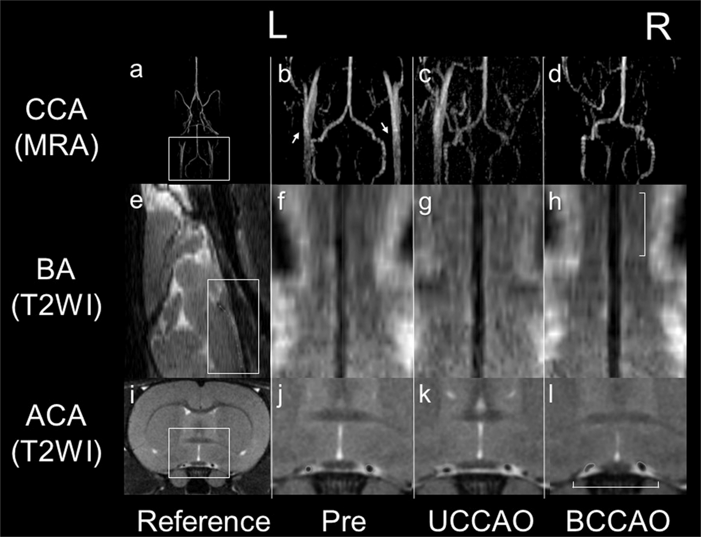
Feasibility Of Ivim Parameters From Diffusion Weighted Imaging At 11 7t Mri For Detecting Ischemic Changes In Common Carotid Artery Occlusion Rats Scientific Reports

Pdf Mr Angiography
Ismrm21 Advanced Liver Imaging Masses Methods
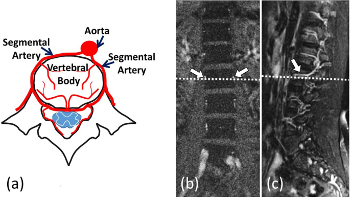
Quantifying Lumbar Vertebral Perfusion By A Tofts Model On Dce Mri Using Segmental Versus Aortic Arterial Input Function Scientific Reports

Emerging Magnetic Resonance Imaging Techniques For Atherosclerosis Imaging Arteriosclerosis Thrombosis And Vascular Biology


0 comments:
Post a Comment